Unraveling forces on cells with artificial microscopic hairs
Jaap den Toonder, Professor of Microsystems, is developing a completely new system to better understand the effect of forces and flows on cells and tissues. He is using a special laser to create artificial cilia, inspired by vibrating hairs that occur in nature. Den Toonder is receiving an Advanced Grant of 3 million euros from the European Research Council (ERC) to carry out this research.
Almost every process in biology, from embryonic development to organ function and the incidence of disease, is based on biomechanical interactions between cells and their environment. If we understand these interactions, we can also better understand, for example, the spread of tumor cells or the brittleness of bones. The forces and flows between cells are often investigated by imitating fluid flows with valves and pumps, but this does not allow you to achieve the precision and control needed to make further steps in this research.
Synchronized motion
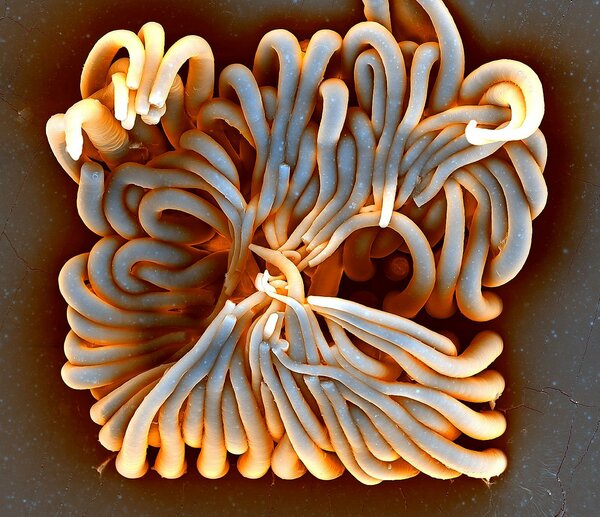
Vibrating hairs inspired Den Toonder to build a new system, with which you can precisely control and study these forces and flows in a laboratory environment. Vibrating hairs, or cilia, are ultra-thin microscopic hairs, which move tightly packed together like a crowd doing the 'wave' in a stadium. Cilia are found everywhere in nature, also in our human body where they play an important role. Their synchronized movement, for example, helps to remove mucus from the lungs and transport eggs from the ovaries to the uterus. By regulating how the fluid flows around an embryo, vibrating hairs even ensure that organs such as the heart develop on the correct side of the body.
Just like real cilia, the artificial hairs must, after an environmental signal, be able to initiate a flow in a fluid or exert mechanical forces on their environment. Then they must be able to detect the forces of reaction from the environment. And all in the same hair. Den Toonder: "The cilia we want to build consist of flexible polymers with magnetic nanoparticles. By activating them with an electromagnet, we can make the hairs move locally exactly as we want them to. This enables us to generate a flow in the surrounding fluid or forces on cells that we grow in the vicinity of the vibrating hairs. We then want to measure the biomechanical response of the cells very accurately."
Glass mold
The cilia that Den Toonder wants to build are only 10 micrometers long and no thicker than 1 micrometer. He also wants to place the hairs very close to each other and give them just the right flexibility to be easily moved by the magnetic fields.
To build them, Den Toonder needs a brand new laser with a small focal point and ultra-short pulses. The laser inscribes very precise structures on a micro scale in a glass plate, which then serves as a mold with which the cilia are formed by means of a casting process. Den Toonder then places these hairs in a so-called microfluidic chip, a piece of plastic with small fluid channels, in which cells and tissues can also be grown.
"We can vary the pattern in which we apply the hairs in the chip. For each biomechanical process we want to study, we make a specific chip. For example, compare it to a CD and CD player. The CD is the chip, it is replaceable. The CD player is our entire system of electromagnet, control and measuring equipment," says Den Toonder.
Besides Jaap den Toonder, Erik Bakkers, professor of Advanced Nanomaterials & Devices, is also a recipient of an ERC Advanced Grant. Read more about his research here.