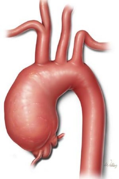
ATAAs, aneurysms in the ascending thoracic aorta, are a life threatening condition. A permanent dilatation of the wall is formed which can range from 3.5 – 10 cm in (maximum diameter). The aneurysms are prone for rupture or dissection. Most patients notice their condition when it’s too late, i.e., when the ATAA has ruptured or dissected. and need emergency surgery which has a high mortality rate.
The standard follow up of ATAAs consists of echocardiographic and CT imaging follow up at regular intervals to identify increase in diameter. It is presently unknown to predict in which patients the aneurysm will dissect or rupture, and whether the root will continue to dilate. The natural history of these roots is poorly known, in part due to the difficult visualization on non-triggered CT’s, due to translational movement of the root. Hence, thorough follow up of any residual native ascending aorta, whether normal in diameter or slightly dilated, is of utmost importance to prevent eventual aortic calamities. Triggered CT’s and 3D echocardiography are the mainstay.
To provide the surgeon with a better understanding of the wall mechanics and an assessment of risk of rupture, biomechanical FE modeling is combined with functional 3D US imaging. The resulting patient-specific models yield not only the stresses in the wall but also the material properties of the wall itself.
Patient study
A multi-center patient study was initiated in the University Hospital in Brussels (UZB), and expanded with the Catherina Hospital Eindhoven (CZE) and the Maastricht University Medical Center (MUMC+). During surgery, regular transesophageal ultrasound imaging is performed. Additional 3-D (+t) ultrasound data are acquired, as well as intraluminal blood pressure. The excised samples are preserved for further testing at our institution.
3D US based FE modeling of ATAAs
Intra-operative 3-D (+t) US data are acquired at the three sites and converted into 3D motion fields and personalized 3D FE models. Mechanical properties resulting from the inverse modeling approach are verified using elastometry techniques, but also by testing the tissue in bi-axial tensile testing devices.
Projects
Projects for bachelor-end projects, internships and MSc projects are available.
• Improve current methods of 3D US based FEM of ATAAs (boundary conditions, loading)
• 4-D functional US imaging of valve, root and aneurysm, elastography of ATAAs
• Quantifying local material properties and non-linearity
• Modeling of interaction between prosthesis and native ATAA tissue post-surgery
• Validation of 3D US techniques with other modalities and/or mechanical testing
Other projects can be designed in consultation with the supervisors (Emiel van Disseldorp & Richard Lopata).