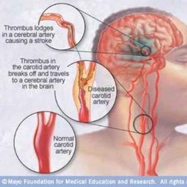
If plaque builds up in the body's arteries, the condition is called atherosclerosis. Over time, plaque hardens and narrows the arteries. This may limit the flow of oxygen-rich blood to organs and other parts of the body. If plaque builds up in the carotid arteries, a stroke can occur.
Currently, intervention planning on carotid plaques relies on the severity of the stenosis, assessed by ultrasound imaging. Plaques are the result of atherosclerosis. The stenosis may limit the flow of oxygen-rich blood to organs and other parts of the body, whereas a vulnerable plaque with lipid content may cause a stroke when ruptured.
The procedure of choice is an endarterectomy. However, only one out of six patients benefits from this intervention. So in practice, the severity of the stenosis is not a reliable criteria for the risk of plaque rupture. Plaque rupture occurs when the mechanical stresses in the cap of the plaque exceed the local tissue strength. Therefore, a biomechanical model of the plaque may help to better assess rupture risk. Ultrasound has the ability to provide highly detailed information on plaque geometry and distensibility. Motion tracking techniques can be used to estimate movement of the wall, strains in the wall and flow in the lumen (see ‘Blood in Motion’).
In our group, new ultrasound techniques, experiments and biomechanical models are developed. In this track, the mechanical properties of the carotid plaques are characterized using ultrasound (PhD Thesis: Renate Boekhoven), but is also pursued using photo-acoustics (Umit Arabul).
Echo-CT Imaging of Vulnerable Plaques
In this project, an experimental framework was established for the mechanical testing of endarterectomy samples, obtained from the local hospital (CZE). These surgical samples contain the intima and parts of the medial layer of the plaque. Testing involves pressurization of the fresh tissue right after surgery, semi-3D ultrasound scanning to obtain geometry, wall motion and strain data and micro-CT imaging to determine the morphology in terms of calcifications and lipid content.
Before applying these methods to real tissue, and in a later stage patients, the ultrasound measurements are also validated and verified in tissue mimicking phantoms (made of polyvinyl alcohol) of straight tubes, stenotic vessels, the latter even including a fatty plaque, and in biological tissue, i.e., porcine carotids obtained at the local slaughterhouse.
Analysis includes ultrasound strain imaging at different angles (Echo-CT), micro-CT scanning, and tensile testing (TT). Both constitutive models can be fit to pressure-diameter (US) or stress-strain curves (TT). Comparison of the two was performed to verify the US results with TT. Micro-CT data was used to build multi-constituent models.
Patient-specific modeling of plaques
In the next phase of this project, these models will be personalized and calibrated using in vitro and in vivo 3-D functional US data. These models cannot only be used to predict patient-specific wall stresses or material properties, but also to verify the strains measured.
Projects
Projects for bachelor-end projects, internships and MSc projects are available.
• Coupling FE models with US strain data
• Registration of US and micro-CT data
• Inverse modeling approach to determine mechanical properties of plaque tissue
• Comparison of in vitro testing of endarterectomy samples to 3-D US data obtained in vivo prior to surgery
• Coupling of strain and flow data in healthy and stenosed vessels
• Construction of realistic plaque phantoms for benchmarking of US techniques
Other projects can be designed in consultation with the supervisors (Richard Lopata & Marcel Rutten).