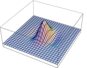
This programme focuses on personalised care for brain tumor patients, and is executed in collaboration with the Department of Neurosurgery of ETZ, with support from NWO TTW (OTP programme), ZonMw (TZO programme), and various academic and industrial partners.
Details: Diffusion weighted imaging (DWI) is a magnetic resonance imaging modality capable of producing scalar images on a six-dimensional ‘(x,q)’-domain. Three of the independent variables refer to position (‘x’) and three to a machine-controllable diffusion gradient magnetic field in the scanner (‘q’). The signal S(x,q) acquired with the help of a suitable diffusion sensitisation protocol can be related to a probability density function P(x,y) for the y-displacement of diffusing (water-bound) hydrogen spins at each position x in the brain, aka the ‘ensemble average propagator’. These high-dimensional images provide a rich source of information about the brain, which can be seen as a porous medium affecting the local diffusivity of water molecules. However, we must then solve the notoriously hard inverse problem to extract quantitative and/or visually meaningful information about the brain’s neural architecture from these complex diffusion data. Our point of departure is to try and relate the physical signals S(x,q) and P(x,y) to mutually conjugate representations of scalar fields that naturally live on the so-called slit-tangent bundle in a Finsler geometric framework, and to exploit the machinery of Finsler geometry for solving the inverse problem. This endeavour is part of the 21st century grand challenge to unravel the human brain, coined ‘connectomics’, which has drawn massive attention worldwide, boosted by the advent of DWI. We are particularly interested in turning scientific results into instruments for improving the neurosurgical workflow.