Brain Image Analysis
Magnetic resonance imaging (MRI) is nowadays extensively used to study structure and function of the brain, e.g. to diagnose and monitor dementia, stroke, and tumors; it is also used to assist in the planning of treatment, e.g. surgical tumor removal, and deep brain stimulation to treat Parkinson.
Magnetic resonance imaging (MRI) is nowadays extensively used to study structure and function of the brain, e.g. to diagnose and monitor dementia, stroke, and tumors; it is also used to assist in the planning of treatment, e.g. surgical tumor removal, and deep brain stimulation to treat Parkinson. Especially of interest is the automatic segmentation and quantification (e.g. volume) of the individual anatomical brain structures and the determination of the structural and functional connectivity between these structures. For example, Hippocampus volume is now recognized as an important biomarker to monitor the presence and progression of Alzheimer disease. Comprehensive visualization of neurological analysis results is a growing research area, including visualization of any uncertainty in these results.
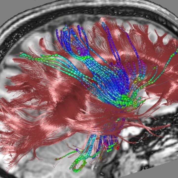
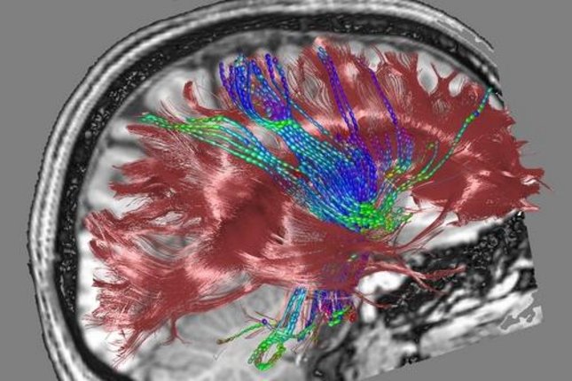
Diffusion weighted imaging (DWI) is the only currently known technique that allows non-invasive visualization of anatomical fibrous structure like white matter in the brain. This magnetic resonance technique allows the measurement of water diffusion characteristics in tissue. The typical size of the fibrous structures is several orders of magnitude below the resolution of clinical scans, and cannot be directly retrieved. However, diffusion in tissue reflects the characteristics of the underlying fibrous structure. This technique has generated an explosion of applications in research given its great potential.