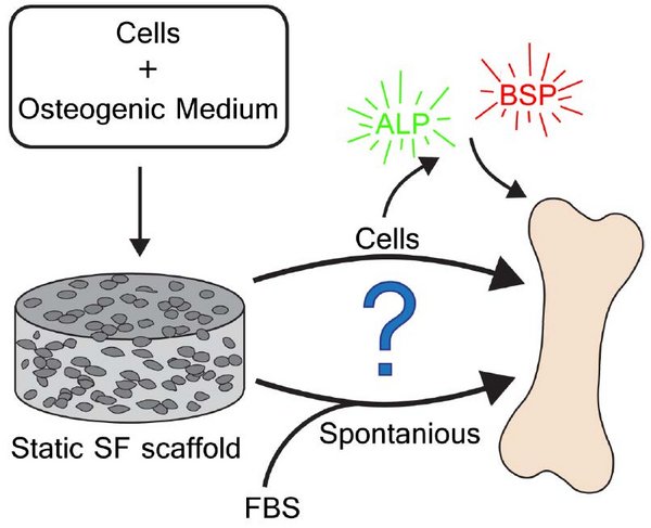
In tissue engineering, silk fibroin (SF) scaffolds have successfully been used for years to support the formation of bone tissue in vitro. Human bone marrow derived stromal cells (hBMSCs) have been seeded on these scaffolds, and under static and dynamic conditions, mineralization has been observed. Studies have shown that hBMSC differentiated towards the osteogenic lineage, did not deposit substantial amounts of extra cellular matrix (ECM) in the form of collagen type-1 (Col I). Instead of the collagen, the SF scaffold started to mineralize. It is still unclear if this was due to a spontaneous process or a cell mediated process. To determine which process caused the mineralization, immunohistochemistry was used for the staining of proteins involved in the process of mineralization. Six week old statically cultured scaffolds with and without cells, were stained for: ALP, BSP, collagen and calcium. ALP and BSP are proteins involved in the cellular process of mineralization. Results showed ALP deposition inside the pores of the scaffold, but ALP and BSP presence inside the SF could not be proven due to autofluorescence of the scaffold. Mineralization of the SF was observed in both cellular and acellular scaffolds thus the process of spontaneous mineralization was confirmed. However, mineralization of the cell-free scaffold was less than the mineralization of the cell-seeded scaffold. It could however not be proven that the difference in mineralization is due to cellular processes. Nonetheless it is believed the cells do have influence on the amount of mineralization as the cell seeded scaffold showed more mineralized SF.