Osteoarthritis (OA) is a degenerative disease of the whole joint, which is highly prevalent and the main cause of disability in the elderly population. Of the large weight-bearing joints in the human body, the knee is one of the most affected joints. During the development of the disease, the morphology of the subchondral bone is altered including trabecular bone sclerosis and an increase of subchondral bone plate porosity. Imaging techniques currently used for clinical investigation of knee OA have a large field of view (FOV) and short scanning times, but a low spatial resolution which limits the investigation of bone microarchitecture. Second generation high resolution peripheral quantitative computed tomography (HR-pQCT) offers the perspective of in vivo assessment of subchondral bone morphology in the knee joint at 61 μm isotropic voxel size. While it is known that trabecular bone morphology can be assessed accurately using second generation HR-pQCT, morphological subchondral bone plate (SBP) parameters have not been investigated. This study aims to determine the feasibility of a clinical study on a population of elderly knee OA patients using HR-pQCT.
First a clinical pilot study was evaluated where two knee OA patients were scanned using the XtremeCT II at 60.7 μm isotropic voxel size. The images obtained were analysed using an evaluation protocol specifically developed for the analysis of the subchondral bone in the knee and morphological bone parameters were determined. Scanning of the knee OA patients brought up a variety of practical and technical issues. Practical issues involved the scanner gantry size limiting the scanning of patients with a larger upper thigh circumference, inconvenience experienced by the patient due to the straining scanning position and relatively long scanning time, and the hard positioning of the knee joint within the FOV. Technical challenges which were encountered were the accuracy of the obtained SBP bone parameters and the suspicion of beam hardening artefacts affecting the obtained images.
The effect of beam hardening and image resolution were investigated using tibial plateaus retrieved from total knee arthroplasty patients (TKA). Osteochondral plugs were drilled from the tibial plateaus at peripheral and central locations within the plateau. To investigate the effect of beam hardening the osteochondral plugs were placed back into the plateau and were scanned with the XtremeCT II (60.7 μm isotropic voxel size) using a custom sample holder. Next, all osteochondral plugs were scanned separately to ensure the absence of beam hardening. Bone mineral density (BMD) of the samples within and outside the plateau was compared. Additionally, the osteochondral samples were imaged using micro CT (VivaCT, 21 μm istropic voxel size) for the assessment of the image resolution of the HR-pQCT images. Bone mineral density (BMD), bone volume fraction (BV/TV) and the subchondral bone plate thickness (SBP.Th) and porosity (SBP.Po) were determined. An underestimation of BMD was found for HR-pQCT imaging of the tibial plateau. The mean whole bone BMD error was 6.9% over all samples and an increase of this error was found in high-density regions, therefore confirming that beam hardening artefacts have influence on the BMD measured in the knee using HR-pQCT. The comparison of the bone parameters of the μCT and HR-pQCT scans showed that image resolution had no influence on the BMD and BV/TV of HR-pQCT images. The investigation of the SBP morphological parameters was inconclusive because of errors made by the custom evaluation protocol used to analyse the images. Refinement of the custom evaluation protocol could provide more accurate results.
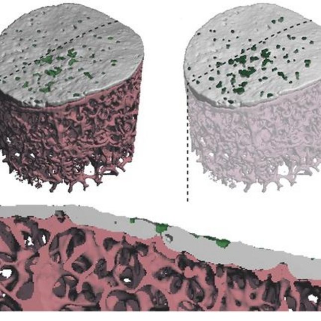
Lastly a detection algorithm was developed to detect perforations in the SBP, which could provide valuable information about the development of knee OA. The algorithm was based on an existing erosion detection algorithm for HR-pQCT scans of finger joints. Perforations are defined by the algorithm as volumes connected to both the outside and inside boundary of the SBP, therefore removing pores which are not penetrating the whole subchondral bone plate. μCT images of the osteochondral samples collected in the previous section were used to assess the functionality of the algorithm and the perforation number (Perf.N), volume (Perf.V) and thickness (Perf.Th) were assessed. A preliminary assessment of the functionality on in vivo HR-pQCT images was done. The detection algorithm was found to be functional for the μCT images and parameters were determined successfully, although errors made by the algorithm were observed. The assessment of the HR-pQCT was less successful since large excess volumes influenced the perforation detection. When the algorithm is refined, in combination with the custom evaluation protocol, it could be a valuable tool for the assessment of the SBP in the knee joint.