Bone fractures are one of the most common orthopedic problems. Among favorable circumstances, a fracture heals spontaneously within two months. However, it is estimated that 5-10% of all fractures is complicated by non-unions, in which natural repair mechanisms remain insufficient. Effective regenerative therapies are still suboptimal for treating critical size bone defects. Therefore, the need for functional tissue substitutes that accelerate bone fracture healing is increasing. This study aimed to develop an in vitro system, consisting of mesenchymal stem cells within a 3D scaffold, that could detect mineralization preferably within two weeks.
Human bone derived mesenchymal stem cells (hBMSCs) were seeded on porous silk fibroin scaffolds and stimulated to differentiate along an osteogenic lineage. The constructs were cultured, either statically, at 60 RPM or at 300 RPM, in spinner flask bioreactors for 28 days. Half of the groups were exposed to 225 nM testosterone and 2 mM alendronate, whereas the others served as control. Immunohistochemistry was used to analyze presence of the androgen receptor. Osteogenesis was evaluated by mCT imaging, histology, scanning electron microscopy, FTIR and confocal fluorescence microscopy.
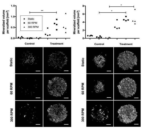
Androgen receptor expression was observed in all constructs after 7 days, which means that testosterone could mediate its effect. Already after two weeks, osteogenic activity and mineralization occurred in each of the constructs, despite culture conditions. Based on micro-CT imaging, significant higher mineralized volumes were however found in the scaffolds that were exposed to the compounds. On the one hand, histological analysis did not show differences in bone matrix deposition between the experimental groups. Alizarin red, on the other hand, indicated that more calcium was present in the treated constructs compared to the controls. Despite, mineralization predominantly occurred in the scaffold material itself.
The results reveal that testosterone and alendronate have a positive influence on bone formation. Nevertheless, it could not be confirmed whether the effect is synergistic, which is consequently the main limitation of this study. The ability of this established 3D system to detect mineralization in silk fibroin constructs after two weeks of in vitro cultivation might be useful for bone tissue engineering applications. It may play an important role in bone fracture repair and may provide an appropriate model to assess the effectiveness of novel drugs.