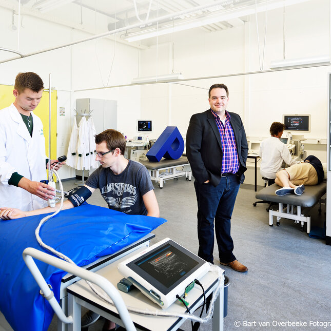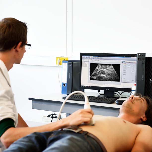
The Photoacoustics & Ultrasound Laboratory Eindhoven, PULS/e, is part of the Cardiovascular Biomechanics group. The PULS/e lab was designed to facilitate the ongoing research on technology development (ultrasound imaging, photoacoustics) and applied science (US-based biomechanical modeling for improved clinical decision making). Ultrasound imaging has always been one of the main imaging modalities in the clinic. Currently, many advances and development in ultrasound imaging are being made.
The lab is fully equipped for development of new functional ultrasound imaging techniques, using both high-end clinical systems with open RF interfaces, but also research platforms, featuring a high-frequency Verasonics system and a fully integrated photoacoustics platform by ESAOTE and Quantel (FULLPHASE).
The lab also hosts advanced experimental mock loops for performing measurements of the mechanical properties of cardiovascular tissue under physiological circumstances and validation of strain imaging techniques, elastography, and vector flow velocity imaging. Moreover, the lab has access to the Laboratory for Biomechanics, where micro-CT and tensile testing equipment are available for validation purposes, and to the Laboratory for Cell & Tissue Engineering and the Microscopy Laboratory for histology. Validation is crucial in getting insight in the performance of new techniques, but also for benchmarking against existing methods, all to improve the acceptation into the clinic.
List of equipment
Video
Contact us
-
dr.ir. Lopata, R.G.P.
-
dr.ir. Maenhout - van Haag, M.
