Researchers grow most lifelike woven bone yet starting from cells
First bone organoid will greatly increase understanding of bone formation and bone diseases.
Researchers from Eindhoven University of Technology and Radboudumc have interwoven various bone cells into an 'organoid' that can independently make new, hard bone tissue. It’s the most complete 3D model of bone formation to date. The 3D model allows for the study of the key biochemical processes in unprecedented detail and could help in cracking the many mysteries surrounding bone formation. Moreover, the lab-grown bone is particularly suitable for testing and designing new treatments for bone diseases such as osteoporosis or osteogenesis imperfecta.
Imagine using stem cells from your bone marrow to grow a new piece of your bone tissue in the lab, after which medical doctors explore how certain drugs affect your bone tissue. In this way, a tailor-made treatment plan could be made for you, and potentially for everyone. Welcome to the world of personalized medicine.
This vision of drug development is no longer science fiction now that researchers from Eindhoven University of Technology and Radboud university medical center have actually realized the first part: growing a lifelike piece of bone tissue from human stem cells. It is the first organoid of bone, a simplified version of the original bone, and the researchers report about it today in the journal Advanced Functional Materials.
Coherent picture
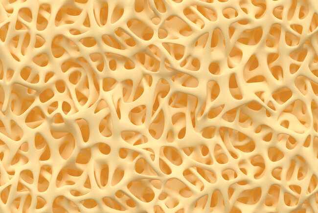
"With this, we present, for the first time, the full picture of early-stage bone formation," says Sandra Hofmann, associate professor in Bioengineering Bone from TU/e. And this is of great importance, particularly as the process by which bones form is still largely a mystery. Bone is a very complex material in which, on the one hand, countless cells and processes interact and, on the other hand, an ingenious matrix of collagen and mineral is built up to provide material strength. Much is known about the individual components, but a coherent picture has been lacking until now.
Three types of cells play the main role in bone formation: osteoblasts (which build bone tissue), osteoclasts (which take bone away) and osteocytes (which regulate the building and breaking down of bone). "Most studies so far have focused on one of these types of cells, but that is not a good representation of the real tissue," says Hofmann. "We present here a piece of woven bone (early-stage bone) that developed from stem cells and contains two types of bone cells: osteoblasts and osteocytes. We now see that we can make lifelike bone exclusively with these two cell types."
Getting wiser from molecular poking
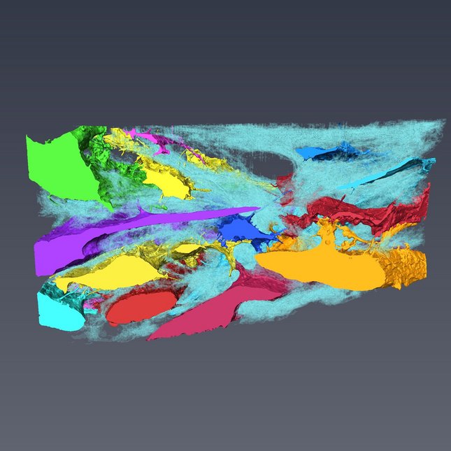
"And perhaps more importantly, our system behaves just like early-stage bone ", says Anat Akiva, assistant professor Cell Biology at Radboudumc. "We show that both types of cells produce the proteins that the cells need for their functionality, and we show with the greatest detail that the matrix actually is the bone matrix we see in real tissue.”
The fact that a simplified representation of the formation of bone at the molecular level is now possible offers unprecedented possibilities, according to the researchers. "A bone consists of 99% collagen and minerals, but there is also another 1% of proteins that are essential for successful bone formation," explains professor Nico Sommerdijk from Radboudumc. "So what’s the role of these proteins? How do they support bone formation? Never before have we been able to look at the milestones of this process at a molecular level."
And with that, they immediately have a great starting point from which to investigate the cause of genetic bone diseases such as 'brittle bone disease' and their possible treatments. "Remember that the origin of many diseases is at the molecular level - and so is the treatment," says Akiva. "In fact, we now have a simple system in a reliable environment where we can poke around and see what happens."
ReferenCE
Anat Akiva et al., An Organoid for Woven Bone, Advanced Functional Materials (9 March 2021). DOI:10.1002/adfm.202010524
A unique and accidental collaboration
Crucial to the creation of the first 'organoid' of bone was the combination of researchers with different backgrounds. "We are one of the first teams that uniquely combines methods from both biology and chemistry, which allows us to look at the first stages of bone formation from the perspective of cells as well as the mineralized collagen matrix," says Hofmann.
“Our different scientific backgrounds created a diverse team of bioengineers, material scientists, and microscopists. This allowed us to think outside the box and to develop our unique approach combining biological and material aspects of bone research,“ says Akiva.
Remarkably, this collaboration originated largely by chance. “For many years I was fascinated by the process of bone formation. After listening to a lecture from Sandra during an annual ICMS symposium at TU/e I knew we must collaborate on this topic,” says Sommerdijk.
"For me, this is the absolute number-one publication," Hofmann says delightedly. "This is what I have been working towards all these years. And at the same time: now that we have come this far, it raises so many new questions that I want to address in future research."