Uterus in motion
PhD student Connie Rees demonstrates that with a renewed ultrasound method, subtle movements of the uterus can be measured. This offers a new perspective on fertility issues in women with endometriosis.
Violent contractions during childbirth, monthly menstrual cramps; we know the uterus can contract forcefully. But the idea of the uterus as a muscle in continuous motion is new. TU/e researcher and gynecologist-in-training Connie Rees uses a renewed ultrasound method to show that these subtle contractions are vital to the proper functioning of the uterus, especially for those with a pregnancy wish. And that offers a new perspective on fertility problems in women with endometriosis. On Tuesday April 2nd, Rees defended her dissertation - with honors - at the Department of Electrical Engineering and Ghent University.
It all started with a personal motivation to be able to diagnose endometriosis more quickly. Because like for many women who suffer from endometrial tissue proliferating inside and outside the uterus, it was an exhausting process for her twin sister to get the right diagnosis, Rees sighs. “Severe pain and heavy bleeding every month; many women suffer in silence and try to soldier through. Or their complaints are brushed aside - “menstrual pain is just part of it.” That really gets on my nerves; endometriosis is an underestimated condition that can seriously disrupt a woman’s life.”
She turned her anger into action and delved into research to increase and spread scientific knowledge about endometriosis. Her main focus was the question of how to diagnose it more quickly and in a less invasive way.
Overlooked
Because that needs to change, Rees emphasizes. “On average, it takes about eight years before a woman with endometriosis is given the right “label’. This is partly due to the female “grin-and-bear-it” mentality and a great deal of misunderstanding, but also because this condition can manifest itself with increasing intensity throughout one’s life. So teenagers or women in their twenties with early symptoms are often overlooked.”
Rees specifically focused on proliferation of endometrial tissue in the uterus itself: adenomyosis. Whereas suspicious endometriosis spots and adhesions in the abdomen are still “relatively” easy to see through laparoscopy, it is much more difficult to get a good look inside the uterus, Rees explains.
“From the outside, there is often nothing to see, and you have to be very experienced to be able to recognize proliferations on an ultrasound. Especially when there are additional conditions that complicate the ultrasound, but also when there are very mild abnormalities. As such, static ultrasound images are difficult to assess afterwards. This is why some hospitals prefer to do an MRI. But a clear protocol of what should be measured and on what criteria adenomyosis is diagnosed is still lacking. Everyone has a different way of doing it.”
Predictive model
So it was high time for a clear approach, Rees and her supervisors decided. Based on an extensive literature review, she identified several factors that can reliably predict the likelihood of adenomyosis. “Both the patient's story and the parameters that we can detect on MRI images are important in this regard. We have created a predictive computational model that can hopefully be used in the clinic soon after a few developmental steps.”
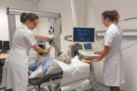
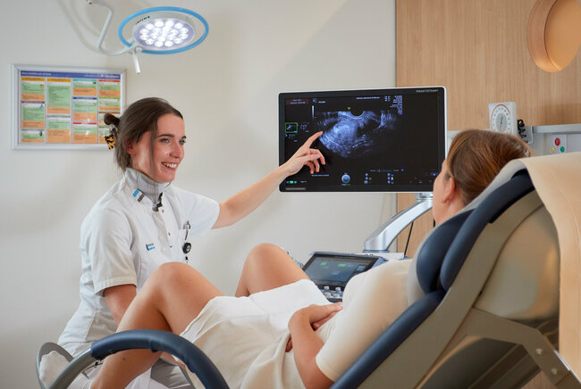
In addition to MRI images, Rees also investigated whether ultrasound methods are suitable for easier detection of adenomyosis. She wanted to further investigate the functioning of an affected uterus as well. For this, she joined in with ongoing TU/e research on visualizing uterine movements. “It was only recently that we learned that the uterus is a muscle in constant motion. But those movements are very subtle, and therefore difficult to measure. With a measurement method that was previously developed at TU/e, it is now possible to objectively monitor uterine movements on ultrasound film. Together with the Catharina Hospital and Ghent University Hospital, we are bridging the gap between technology and practice.”
Modified IVF treatment
Using this renewed ultrasound method, Rees and her colleagues observed how the uterus moves in healthy women, and soon, Rees switched to women with adenomyosis. Where she would normally see organized movements, she was shocked by what she observed in affected uteruses.
“A state of chaos. A normally functioning uterus is most active around ovulation. That’s when a sperm cell needs to be taken to the fallopian tube in order to fertilize the egg. And when the fertilized egg starts nestling in the uterine lining, the movements are supposed to be calmer. Adenomyosis causes disturbances of the uterine muscle; we have observed abnormalities in two important phases of the female cycle. During ovulation, there is much less movement; besides, the movements go in all directions. This can make it much more difficult for a sperm cell to reach a mature egg in time, or at all. And in a later phase, when a potential egg is supposed to start nestling, the uterus is actually more active.”
Early diagnosis
Early diagnosis of adenomyosis or endometriosis is extremely important for young women, Rees stresses. “If we can see on ultrasound film that the uterine movement is disrupted, we can start taking that into account for follow-up steps. We could try to normalize uterine motion with medication. And we could then also use the new ultrasound method to monitor whether the medication is effective. If natural conception is unsuccessful, we could opt for a modified IVF treatment based on the uterine contractility.”
PhD in the picture
What is that on the cover of your dissertation?
“For this, too, I decided to use new technology: I created the front cover using DALL-E. At first glance, it appears to be flames, but if you look more closely, you can see a uterus with disrupted movements.”
You’re at a birthday party. How do you explain your research in one sentence?
I always start by asking if you know what endometriosis is. Fortunately, more and more people are responding with a “yes” - though it’s often followed by harrowing stories. There has been a real change in recent years, thanks also to all the attention in the media. I research how uterine disruption affects fertility and pregnancy.”
How do you blow off steam outside of your research?
“As a physician-researcher, I shuttle back and forth between the research facility, delivery room and consultation office. Between TU/e, Catharina and Belgium. That leaves little time for hobbies; watching Netflix on the couch is how I unwind in the evening. Fortunately, I derive tremendous satisfaction and energy from supervising students. Through them, I can also expand knowledge about endometriosis.”
What tip would you have liked to receive as a beginning PhD candidate?
“Build a professional network and seek out collaborations, it will really advance your research.”
What is your next step?
“I loved the combination of science and working in the clinic. As a physician, you stay in touch with the people you’re doing it for, and as a researcher, you don’t lose sight of practice. I hope to complete my gynecology program in Ghent in a few years and would then like to specialize in endometriosis. So that hopefully, in the future, people like my sister won’t have to wait so long for their diagnosis.”
Large study
As part of the large-scale retrospective ADENO study she subsequently set up - the first of its kind worldwide - Rees examined 8,000 pregnant women with adenomyosis to see if there might also be a link between their condition and pregnancy complications. “It turns out that women with adenomyosis run a higher risk of experiencing high blood pressure and preeclampsia or having to undergo an (emergency) cesarean section during pregnancy. So here too, early diagnosis is important for proper medical management during pregnancy and childbirth.”
Under the rug
Rees expects that uterine movement research will lead to many innovations within the field of gynecology. “The field is slowly gaining momentum; we have taken important first steps. It would be great if we could diagnose certain conditions such as endometriosis and adenomyosis in an early stage using an ultrasound that feels normal to the patient. And it works both ways: in the case of fertility problems, it can be used as a diagnostic tool to visualize any disturbances. I want to make this technique as accessible as possible. Because when it comes to gynecological conditions, things have been swept under the rug for far too long.”
More information
Learn more about the work of Connie Rees in this article from Catharina Ziekenhuis.
The PhD research by Connie Rees is part of the Eindhoven MedTech Innovation Center (e/MTIC) and in collaboration with Catharina Ziekenhuis.
Source: Cursor (door Nicole Testerink)
More on Health