Less invasive for patients: using blood tests to diagnose lung cancer
Sylvia Roovers-Genet defended her PhD thesis at the Department of Biomedical Engineering on February 1st.
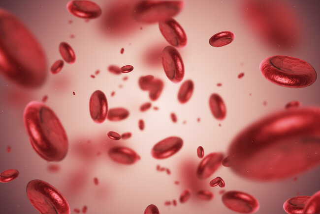
Currently, when lung cancer is suspected, a ‘morsel’ of tissue is removed and examined under the microscope. This may change in the future. During her PhD research, Sylvia Roovers-Genet examined proteins in the blood of people with, without, and with possible lung cancer, and thereby developed a method to demonstrate the presence of lung cancer through blood tests. This method can be developed in the future, with the goal of making it suitable for predicting lung cancer.
Even at the start of her PhD at the Department of Biomedical Engineering, Sylvia Roovers-Genet knew that she would be deliberately working towards a clinical application. Not surprising either when you consider that she conducted her research in close collaboration with colleagues at the Catharina Hospital and the Máxima Medical Center in Eindhoven/Veldhoven. The goal was to look for characteristic proteins (markers) in the blood of lung cancer patients.

Roovers: “It is important to know that people often talk about lung cancer as if it were one disease, when in fact there are many different forms. Each with its own treatment and approach. So, for a lung specialist, it is particularly important to know exactly which variant he or she is dealing with.”
Current approach
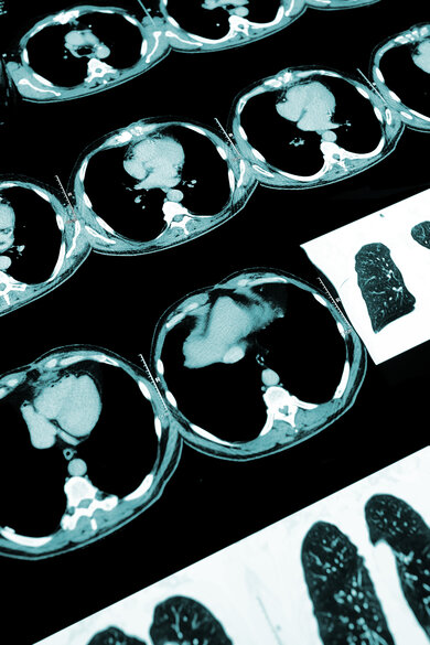
“Currently, there is a gold standard for determining whether someone has lung cancer. If suspected, the first step is a scan, such as CT or PET CT. That gives insight into where the symptoms may be coming from and the location of possible cancer cells or a tumor."
The second step is a biopsy. “To be sure that it is cancer and not some other condition, a biopsy of lung tissue is now the only way to provide certainty. But that evidence of tumor cells cannot always be obtained. Sometimes people are too old or too sick and the biopsy itself is too risky for their health. And sometimes people refuse to undergo the procedure.”
Looking for a new method
“That’s why we started looking for a method that is much less invasive for patients. Blood sampling is much less painful and risky. Especially for the high-risk lung cancer group, where smoking and age are the main risk factors,” Roovers says.
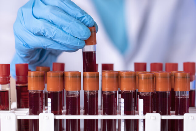
For the study, it was important to at least examine blood from three groups of people: with, without, and with possible lung cancer. “As many as six Dutch hospitals cooperated in the study by asking patients if they wanted to participate when they came to the pulmonologist with suspected lung cancer. Relevant patient data were then recorded and their blood was examined to ascertain the proteins.”
In the end, more than a thousand people participated in the study. Because of the size of that group, the results from Roovers’ doctoral research are very reliable. For example, it was interesting to see that in thirteen percent of patients, the current gold standard could not conclusively determine whether or not these patients had lung cancer. For these patients, blood tests might offer a solution in the future.
Protein analysis to demonstrate lung cancer
The patient survey provided a huge database that Roovers and fellow researchers were able to work with. “We developed new detection methods for quantifying two promising lung cancer protein tumor markers based on liquid chromatography-tandem mass spectrometry (LC-MS/MS). This is a method that looks at the weight of proteins in the blood and thus recognizes tumor markers.”
“This allowed us to detect these markers even at very low concentrations in the blood of patients with lung cancer. The new methods can help in follow-up studies to find out whether LC-MS/MS-based detection can also add value in clinical practice in the hospital compared to current methods,” says Roovers.
Step closer to the GP surgery
To facilitate the step to the hospital, or clinical practice, Roovers and colleagues developed not only the diagnostic method but also a decision algorithm for GPs. “It’s not one marker that you have to pay attention to as a GP but a combination of factors. That’s why we help GPs correctly interpret blood test results according to our method. We can now use our method, based on blood tests, to be able to say with at least 95 percent certainty for two-thirds of patients that they have lung cancer.”
Asked about the practical application, Roovers still exercises restraint. “We have now scientifically proven that lung cancer can be demonstrated in blood. The next step is always a validation study in practice. That is being set up and taken up by a collaboration of TU/e, Catharina Hospital and Máxima Medical Center.”
A jab at the GP surgery?
And after that? Just go to the doctor and get a blood test to find out whether you have lung cancer? “Who knows,” Roovers smiles. “But it's not that far off for now. Make no mistake, lung cancer is unfortunately still very deadly. After five years, only 19 percent of people with lung cancer are still alive. So, it would be great to detect precisely these forms of cancer earlier, so they can be better treated.”
Title of Sylvia Roovers-Genet's dissertation: Lung cancer protein tumor markers: Development, validation and clinical use of quantitative serum tumor marker assays.
Supervisors: Volkher Scharnhorst and Luc Brunsveld
More on Health

Latest news


![[Translate to English:]](https://assets.w3.tue.nl/w/fileadmin/_processed_/e/0/csm_BvOF%202019_1031_BHF%20license%20TUe%20ILI%20copy_8a50884392.jpg "[Translate to English:]")