Improved monitoring of dangerous aneurysms
During her doctoral research, Esther Maas investigated the use of new ultrasound techniques to image dangerous aortic aneurysms for patient-specific care.
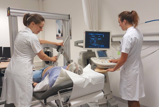
An aneurysm is a dangerous vascular condition in which a weak spot in a vessel wall can bulge like a balloon. In the aorta, the largest artery in the abdomen, such an aneurysm is life-threatening, especially when it bursts. During her doctoral research, Esther Maas worked on a reliable way to monitor aneurysms using ultrasound. She conducted her research at the TU/e PULS/e group (Photoacoustics & Ultrasound Laboratory Eindhoven) and the Catharina Hospital in Eindhoven. She defended her thesis at the Department of Biomedical Engineering on April 9.
For such a dangerous vascular condition, an aneurysm is often discovered late or even too late. In fact, an aneurysm itself almost never causes any symptoms. It is only when the bulge bursts and bleeding occurs that an aneurysm is usually discovered afterward. When this happens in our body’s most vital artery, the aorta, it is life-threatening.

Tracking an aneurysm
Sometimes aneurysms are discovered earlier anyway, usually by chance. For example, during a scan of the abdominal cavity because of other symptoms. In that case, the vascular surgeon can continue to monitor the bulge and intervene when the risk of the aneurysm bursting becomes too great.
“The probability of an aneurysm rupturing is related to its size. Yet we also see differences in patients,” Esther Maas explains. “Sometimes relatively small aneurysms also rupture, while in other patients a relatively large aneurysm remains stable.”
Diameter and shape
“The current criteria for operating to eliminate the aneurysm are mainly related to diameter, which means that sometimes surgery is performed too soon and sometimes too late. We wanted to improve that by improving our understanding of when an aneurysm is at risk of rupturing. To do this, we looked at the three-dimensional shape of the aneurysm and its mechanical properties.”
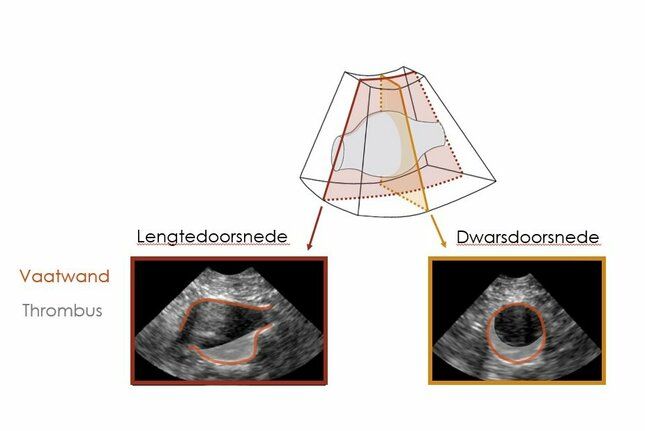
Currently, standard two-dimensional ultrasound is employed to monitor an aneurysm. The disadvantage of this technique is that a picture can only be taken at one location at a time (‘a slice’). While MRI and CT allow the entire three-dimensional shape of the aneurysm to be viewed, these techniques are expensive and time-consuming.
RELIABLE ultrasound
So Maas took up the challenge of using ultrasound to develop a reliable method of monitoring aneurysms. So that patients can be monitored safely, healthcare costs saved and doctors spared the task of operating unnecessarily.
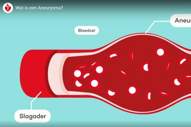
What is an aneurysm?
An aneurysm can occur in any artery but is most common in the aorta (the large body artery). And then usually in the abdomen. An aneurysm is life-threatening.
What is an aneurysm?
An aneurysm is a localized dilation or bulge of a blood vessel. The diameter of the blood vessel is then more than one and a half times larger than normal. Watch the video (in Dutch, ed.) to the left to learn more about an aneurysm. For example, about the symptoms, consequences, and treatment.
3D ultrasound over time
3D ultrasound, especially as moving images (temporal 3D or 3D+t-echography), is an excellent method for imaging an aneurysm. It is fast, less stressful for the patient, does not emit radiation, and is also more affordable than the alternatives. Maas: “It allows us to see the shape and movement of the aneurysm simultaneously.”
The image quality could still be improved to allow the doctor to assess an aneurysm. “Processing these ultrasound images is challenging, however, because the images contain a speckled pattern. And, in addition, the gray shades are not directly traceable to a tissue,” Maas says.
Determining shape automatically
Maas: “Therefore, at the beginning of my research, we mainly worked on a method to automatically and accurately determine the shape of an aneurysm from 3D+t ultrasound images.”
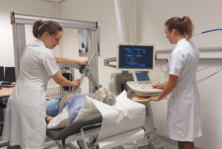
The actual patient images were taken at the Catharina Hospital in Eindhoven. As many as five hundred patients were monitored in this study. Maas: “When they came for a conventional two-dimensional ultrasound for diameter control, we also made a 3D+t-echo.”
“We developed a computer algorithm that looked for the vessel wall in the 3D+t-echo images. We compared the resulting shape to CT (computed tomography) images, the current gold standard for imaging aneurysms. This showed that the shape from 3D+t-echo images corresponds well with the shape from CT.”

Predicting growth of aneurysms
By automatically determining the aneurysm shape on all images of the 3D+t ultrasound time series, it was also possible to estimate the expansion of the vessel wall during the cardiac cycle. Combined with blood pressure, this gives an estimate of how elastic the vessel wall is.
“Using this technique, we found out that a combination of three-dimensional shape and elasticity has a better predictive value for aneurysm growth than measuring diameter alone,” Maas continues.
“In the next step, we further improved the shape determination using deep learning. We used over 1,300 available 3D+t ultrasound images to train a model that could even more robustly determine the shape of the aneurysm.”
Improve imaging
But the researchers wanted to improve imaging even more. By combining multiple ultrasound images, they wanted to solve some of the shortcomings of ultrasound, one of which is that only a small area can be viewed at a time.
“This meant that the ultrasound image did not capture large aneurysms in their entirety,” Maas explains. “To solve this, we took multiple ultrasound images side by side, first synchronizing these in time and then spatially superimposing and fusing them. This allowed us to image even larger aneurysms with ultrasound.”
Differences in direction
A second disadvantage of ultrasound is directional – the direction in which you look is very decisive. In one direction the image quality is much better than in the other direction.
Maas: “To solve this, we combined ultrasound images of the aorta from different angles, which led to an improvement in contrast in the images and a better determination of expansion during the cardiac cycle.”
Big step in aneurysm care
“Our research showed how aneurysms can be monitored using 3D+t-echo images,” Maas concludes. “First, by automatically determining different characteristics that are important for the growth of aneurysms in the abdominal cavity. In addition, by demonstrating the added value of combining multiple ultrasound images.”
This marks a major step towards being able to use 3D+t ultrasound images to monitor aneurysms in the clinic, ultimately providing more patient-specific care.
Esther Maas defended her dissertation "Characterization of abdominal aortic aneurysms using time-resolved 3D ultrasound" at the Department of Biomedical Engineering on April 9, 2024.
Promoters: Richard Lopata and Marc van Sambeek
The research was conducted at Eindhoven University of Technology and Catharina Hospital Eindhoven and made possible in part by funding from ERC Horizon 2020.
Esther Maas’ doctoral research is part of the Eindhoven MedTech Innovation Center (e/MTIC).
More on Health
Latest news