Determining MRI scanning restrictions for patients with medical implants
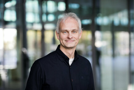
Peter Stijnman defended his PhD thesis at the department of Biomedical Engineering on June 30th.
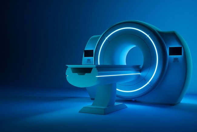
In general, patients with medical implants cannot undergo MRI examinations as the magnetic fields used by the MRI to create an image can interact with implants, resulting in a variety of potential safety hazards, such as forces, vibrations, and tissue heating. The latter is difficult to quantify because it depends on a number of factors such as patient anatomy and location of the implant. Simulations can help with these decisions on MRI imaging, but current simulations take some time. For his PhD research, Peter Stijnman developed a new, faster simulation approach that drastically speeds up tissue heating evaluation.
Before a patient with an implant undergoes a magnetic resonance imaging (MRI) examination, a risk versus benefit is made for the patient. However, the risk of tissue heating due to the interaction of the radiofrequency field from the MRI scanner with the implant is very difficult to quantify. Even if the patient does get an MRI scan, multiple timeslots are reserved or images with lower quality are made, limiting the diagnostic value.
Difficult to quantify
Added to this, the potential heating of medical implants is difficult to quantify because it depends on a number of variables. These include the MRI scanner, the patient anatomy, the implant geometry, and the location of these three with respect to each other.
As a result, measurements and simulations of the potential heating near a specific medical implant are combined to find a worst-case scenario. Based on this worst-case scenario, safe scanner restrictions are set for all patients with that specific type of implant. However, these worst-case scenario restrictions are often overly conservative for most people in a particular patient group.
Faster simulation
For his PhD-research, Peter Stijnman and his collaborators from the Medical Image Analysis group have developed a new simulation method that is much faster compared to the simulation methods currently used.
Rather than waiting hours for one simulation to finish, it’s now possible with Stijnman’s new simulation to obtain results within one or two minutes. The main reason for this speed-up is that the simulation makes use of existing simulated field distributions where the alteration to this field by the implants is calculated efficiently as a perturbation to the existing field.
Using the new simulation method, it is possible to determine patient-specific scanner restrictions rather than ones based on an overall worst-case scenario. A potential workflow to obtain these patient-specific restrictions is also demonstrated in Stijnman’s research. This workflow uses X-ray images of the patient made after the medical implant was placed to determine the geometry and location of the medical implant.
The benefit of using patient-specific scanning restrictions is that either a smaller timeslot needs to be allocated for that patient or, in the same timeslot, an image with higher quality can be obtained. Furthermore, for some patients, the worst-case restrictions might be prohibitively strict, while a patient-specific approach would allow them to have an MRI exam.
Title of PhD-thesis: “MRI safety of implants: measurement and model-based tools for validation and calculation of RF field interactions.” Supervisors: Josien Pluim (TU/e), Cornelis van den Berg (UMC Utrecht). Co-supervisor: Alexander Raaijmakers (TU/e) . Other main parties involved: UMC Utrecht Utrecht, Philips Best, Medtronic Minneapolis
Media contact
More on Health
Latest news


![[Translate to English:]](https://assets.w3.tue.nl/w/fileadmin/_processed_/e/0/csm_BvOF%202019_1031_BHF%20license%20TUe%20ILI%20copy_8a50884392.jpg "[Translate to English:]")