New imaging approaches to analyze carotid artery plaques
Jan-Willem Muller defended his PhD thesis at the Department of Biomedical Engineering on March 8th.
Stroke is the second cause of death and the third cause of disability worldwide. It is also a difficult disease to prevent and to cure. A significant proportion of all strokes is caused by carotid artery disease, in which so-called plaques are growing on the inner side of the carotid artery. The plaques can be hard to see and analyze with non-invasive methods in clinical practice. That’s why Jan-Willem Muller researched two main methods of medical imaging, ultrasound (US) and photoacoustic (PA) imaging of the carotid artery. Additionally, he evaluated their reliability in predicting dangerous plaques and the risks they pose for a stroke.
A stroke is caused by a sudden interruption of blood supply in the brain and often leads to permanent brain damage. A major cause of stroke is the rupture of atherosclerotic plaques in the carotid artery. That means it is imperative to detect these plaques early on and remove them when they could potentially cause a stroke.
According to the current medical guidelines surgical treatment should be considered for patients with a plaque-caused obstruction of the carotid artery of over 50%. This will be usually measured using ultrasound (US) imaging.
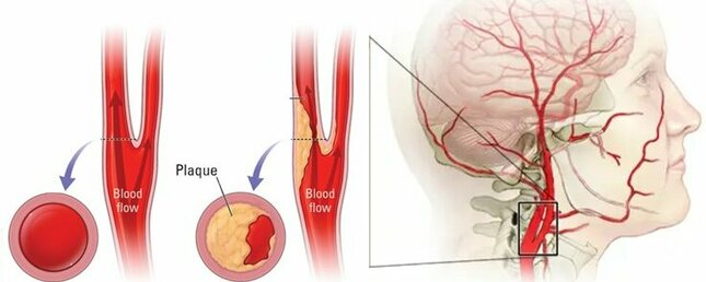
Unfortunately, this guideline leads to the overtreatment of patients. This in turn leads to unnecessary surgical risks and also puts an additional burden on healthcare systems worldwide. Nowadays, that means we are operating on 10-20 patients to prevent one stroke. Therefore, a more appropriate and patient-specific indication for preventive surgery to remove the plaques from the carotid artery (carotid endarterectomy) is needed.
Evaluating currently available imaging techniques
Current ultrasound techniques used in a clinical situation are only able to estimate the size and percentage of the plaque obstruction. Unfortunately, they cannot differentiate sufficiently between stable plaques and plaques prone to rupture. Only the latter pose a serious stroke risk and should be surgically removed.
Carotid photoacoustic (PA) imaging and strain imaging are harmless preclinical methods that extend traditional ultrasound imaging. Adding them to the imaging protocol could provide the required additional optical and mechanical information. This should allow the physicians to detect which plaques should be removed.
In PA imaging, a short laser pulse is used to generate an acoustic response, which can be sensed using ordinary clinical ultrasound transducers. By analyzing this response for many different optical wavelengths (colors), the material types present in the plaque can be determined.
Verifying the imaging results
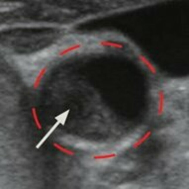
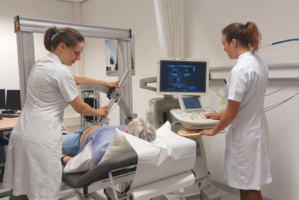
In his research, Muller verified the ability to image carotid artery plaques in humans in an intraoperative clinical pilot study in which the carotid artery was exposed. The intraoperative scanning successfully yielded the first PA images of human carotid plaques.
During the procedure, a unique hand-held probe was used integrating an ultrasound transducer and two laser-diodes to acquire the images. After analyzing the results Muller concluded that strong signals could be observed in human plaques containing hemorrhages. Hemorrhages are predictors or precursors to an actual rupture in the plaque.
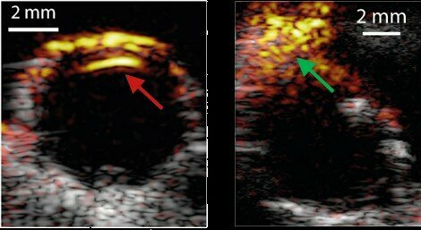
The two laser wavelengths used were sufficient to separate signals imaging the plaque from those generated by the blood flow inside the artery (blood pool in the lumen). Still, more wavelengths are needed for a more detailed analysis. The method is therefore shown to work in human plaques, although more research is needed to improve diagnostic value,” recommends Muller.
Mechanical characterization
Besides PA imaging, Muller used mechanical characterization to analyze the soft tissue composition in carotid plaques. The mechanical characterization was performed by measuring strain values within the plaque. This information can be used to provide information on the nature of the tissue present in the plaque. For example, fatty tissue is soft and prone to rupture and fibrous tissue is hard and more stable. The strains can be determined from measured displacements in the plaque using motion tracking.
To improve the motion tracking results and avoid unphysical displacement fields, a mathematical method was developed.
The developed US imaging and mechanical characterization techniques were applied to porcine blood vessels and human carotid arteries. The method improves the consistency of the measured displacement fields using US images and reduces accumulated errors in high frame rate acquisitions. The improved estimation of the displacement fields can be used to increase the mechanical characterization accuracy of the plaque’s soft tissues. This method may therefore lead to improved clinical mechanical characterization of the plaque and support decision making.
Combining modeling and imaging to improve results
Unfortunately, a common problem in both PA and US strain imaging modalities is the lack of a solid benchmark in human data, which complicates the interpretation of the data. In contrast with experiments, computer modeling tools offer a platform to perform digital imaging experiments in which all relevant parameters are known and fully controllable.
Computer modelling therefore plays an important role in the validation of preclinical methods and facilitates the development of novel methodologies. The current models generally lack realism and are not able to incorporate all relevant acoustic imaging artifacts.
To further improve the realism of computer-generated images in both photoacoustic imaging and strain imaging, novel methods have been developed by Muller. These methods significantly improved the realism of the simulated images and may therefore be used to accelerate future research for both imaging methods.
Title of Jan-Willem Muller’s dissertation: Photoacoustic and elastographic methods for carotid plaque characterization.
Supervisors: Richard Lopata and Marc van Sambeek
More on Health
Latest news
![[Translate to English:]](https://assets.w3.tue.nl/w/fileadmin/_processed_/e/0/csm_BvOF%202019_1031_BHF%20license%20TUe%20ILI%20copy_8a50884392.jpg "[Translate to English:]")