Expert-level automatic techniques for early detection of esophageal cancer

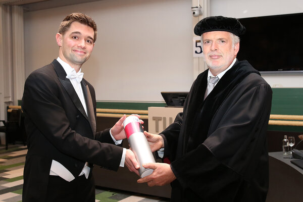
The detection of the early stages of esophageal cancer in Barrett’s Esophagus, based on visual endoscopic detection and biopsies, yields poor detection rates. The PhD-research of Fons van der Sommen presents several new, automatic detection techniques based on digital images provided by endoscopic inspection of the esophagus. The newly developed techniques are almost as good as the top specialists in the field in recognizing signs of early cancer, and in one case even better. The results are an important step towards real-time high quality computer-aided detection (CAD) of cancerous lesions during endoscopic inspections. Yesterday, Van der Sommen gained his PhD with distinction.
Barrett’s Esophagus (BE) is a medical condition in which the body replaces the normal lining of the esophagus with acid-resistant cells in response to reflux. As BE leads to a 30-fold increased chance of developing esophageal cancer, patients regularly undergo endoscopic inspection. When detected early, esophageal cancer be can treated instantly, endoscopically. If detection happens at a later stage, the treatment involves drastic surgery and radiochemotherapy, and the prognosis is severely worse. Better detection techniques for early cancerous lesions are therefore much-wanted, not only to avoid patient suffering, but also to reduce the cost arising from extensive treatment. Providing such techniques is the aim of this research.
The first part of the research focused on automatic detection based on images provided by regular, white-light endoscopy. The visual properties of the developing cancerous lesions were examined and captured with quantitative image features that were fed into algorithms for a CAD system. This system is almost as good as four international experts on Barrett’s cancer in recognizing the presence of cancer in endoscopic images. However, the algorithms tend to draw tighter boundaries of the cancerous tissue than the human experts do.
A separate algorithm was developed for preprocessing of images, in order to automatically exclude images that are not of sufficient quality, e.g. not sharp, with too much reflection, etc. This prevents false positives based on faulty images and hence increases the reliability of the CAD.
The research also looked into a relatively new endoscopic technique, Volumetric Laser Endoscopy (VLE). This technique uses near-infrared laser light to scan the esophagus tissue up to a depth of 3 millimeters. As cancerous lesions arise below the surface, this technique could provide much better early detection, especially of squamous-cell carcinoma, the most common type of esophageal cancer in the Western world. Unfortunately the images generated are complex and noisy and therefore hard to interpret by the human eye. To tackle this problem, CAD algorithms were developed. These clearly outperformed trained VLE-experts in the detection of early cancer.
The initiative for this research came from doctor Erik Schoon, BE-specialist at the Catharina Hospital in Eindhoven. He figured that if smartphones can recognize faces, then it must be possible to learn computers to detect cancer. He contacted Peter de With, professor of Video Coding & Architectures, with this question, and Van der Sommens work is one of the results. The research was done in close cooperation with the Catharina Ziekenhuis in Eindhoven.