Measuring tissue perfusion is valuable for diagnosis and treatment monitoring in numerous diseases. The first part of the perfusion study (Maarten Heres) is to examine the feasibility of Photo-acoustic (PA) imaging as a tool for quantitative measurements on tissue perfusion. Power Doppler (PD) ultrasound is used to benchmark PA imaging. The second track of this study is the assessment of skeletal muscle perfusion during exercise, using PD ultrasound.
Photoacoustic imaging: looking at skin perfusion
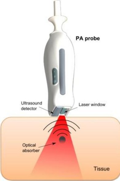
This technique is based on the absorption of short laser pulses in tissue, which results in the emission of ultrasonic pulses. These pulses are detected with a conventional ultrasound (US) system at the surface of the tissue and used for imaging or tissue characterization. In the near-infrared region, hemoglobin is a strong absorber of the light energy. This makes PA imaging very sensitive to the presence of blood.
Approach
• Construction of perfusion phantoms. Different phantoms are constructed, cylindrical vessel phantom with a large luminal area, and solid tissue phantoms that contain micro-channels, resulting in a known perfused area ratio. The phantoms are constructed to mimic (photo)acoustical tissue properties, i.e., have realistic acoustic and absorption characteristics, and are thus suitable for both PA and US imaging.
• Pre-clinical validation. Measurements in superficial tissue are conducted such as the skin and skeletal muscles in the arm and leg. Perfusion is altered minimally invasive by means of temporary occlusion of the supplying artery or exercise. Photo-acoustic imaging data are compared to other techniques, such as Power Doppler ultrasound and near-infra-red spectroscopy.
• Patient studies. Ultimately, the new PA based technique will be used in patients with muscular atrophy (in collaboration with University Medical Center Utrecht), localized scleroderma (in collaboration with Radboud Medical Center Nijmegen) and decubitus (collaboration with STBE group).
Power Doppler Ultrasound: measuring muscle perfusion
This technique is very sensitive for the detection of moving blood in deeper lying tissue, such as the skeletal muscles. In cooperation with the Maxima Medical Centre in Eindhoven, a project is started in which Power Doppler Ultrasound is used to measure the changes in muscle perfusion during exercise. The technical question is whether PD is a feasible and valid tool for the tracking of muscle perfusion. The clinical question that we want to answer is whether the muscle perfusion might be the limiting factor in exercise tolerance for heart failure patients.
Approach
• Development of setups and protocols to measure muscle perfusion during exercise. In the PULSe lab, professional sports equipment is available to perform realistic, (high intensity) exercise measurements in a controlled setting. PD scanning during exercise is possible by means of smart tools that fixes the ultrasound probe to the human body.
• Development of algorithms and models to analyze and quantify the obtained Power Doppler signals. With this data, we can track the kinetics of muscle perfusion and relate them to the exercise intensity.
• Volunteer and patient studies. Reproducibility and validity of our methods will be tested in a volunteer study. Patient studies will be designed based on the results of the volunteer studies.
Projects
Projects for bachelor-end projects, internships and MSc projects are available.
• Photo-acoustic imaging of skin disease
• Oxygen saturation assessment with photoacoustics
• In vivo perfusion measurements in the extremities (arms, legs) during exercise.
• Coupled muscle perfusion and deformation measurements in the upper leg and calf
Other projects can be designed in consultation with the supervisors (Maarten Heres, Richard Lopata).