Imaging
Aim in the track ‘Imaging’ is to develop clinical measurement techniques to assess the geometrical, biomechanical and morphological properties of primarily cardiovascular tissue (heart, arteries/veins), and in the musculoskeletal system, i.e., muscle, cartilage, and skin.
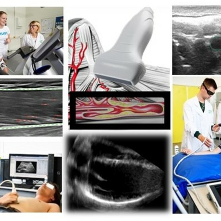

Aim in the track 'Imaging' is to develop clinical measurement techniques to assess the geometrical, biomechanical and morphological properties of primarily cardiovascular tissue (heart, arteries/veins), and in the musculoskeletal system, i.e., muscle, cartilage, and skin. The ultimate goal is to improve diagnosis, clinical decision making, and monitoring of disease progression over time, and/or guide therapy/intervention using a non-invasive imaging modality that is easily accessible, cost-effective and radiation-free.
Focus is on ultrasound imaging, which combines a high spatial and temporal resolution and enables functional imaging. New 3-D and 4-D ultrasound techniques are developed for improved imaging of these organs, but also for functional measurements of motion/flow, strain and biomechanical parameters. Moreover, molecular imaging is performed by means of non-invasive, spectroscopic photoacoustic imaging to differentiate between different tissues based on their optical absorption properties. Use of ultrasound under dynamic conditions has been developed for imaging during exercise in the fields of cardiology, musculoskeletal research, and sports medicine. A large area of research is patient-specific biomechanical modeling of organs using the aforementioned imaging data as input.
Techniques are validated experimentally and pre-clinically to assess its applicability and merit before being translated into the clinic. Many clinical collaborations are established for 'first in man' and/or feasibility studies, as well as clinical pilot studies and longitudinal studies in vivo.