Purpose
Osteoarthritis (OA) is a progressive degenerative joint disease, which is one of the most prevalent causes of joint pain and the leading cause of disability and morbidity. With the knowledge that OA is progressive and difficult to treat, early diagnosis and close monitoring is desirable. To this end, a qualitative arthroscopic tool could be profitable for multiple purposes. Ultrasound (US) is a promising technique to meet this demand due to its high resolution, ability to evaluate soft tissue directly and potential capability of early cartilage damage detection. This study first aims to show the potential of ultrasound to detect cartilage damage by morphological features rather than surface measurements. The second aim was to perform US elastography to detect locations of high strains in articular cartilage after mechanical overloading. The final aim was to investigate histology techniques for cartilage damage detection after mechanical indentation to correlate this damage to the structural differences seen in US.
Materials and methods
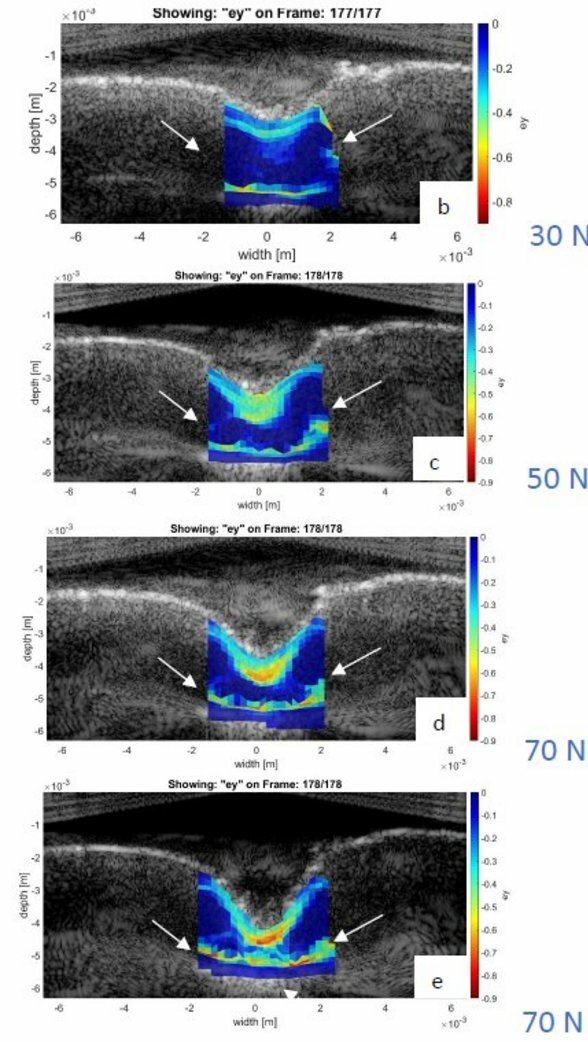
Tibia plateaus obtained from goats that were treated with various osteochondral implants were imaged with a high-frequency probe of 31.25 MHz. The images were processed with a quantitative algorithm, evaluated by two raters and compared to the Ultrasound Roughness index and histological pictures after Indian ink staining. Then, US elastography was performed on bovine patellas. The cartilage was subjected to a mechanical load series of 30N, 50N, 70N and a second 70N for 15min. In between the indentation loading, the recovery of the cartilage was acquired with US for 30 min at a framerate of 0.5 Hz. The US speckle pattern was tracked over time to calculate estimated strain maps in 2D. Finally, cartilage tissue was mechanically loaded to damage it and then examined with different histological staining methods to detect compositional and structural changes as seen with US.
Results
Cartilage on implant-worn tibia plateaus could be distinguished from cartilage on healthy tibia plateaus by US evaluation. As expected, the Ultrasound Roughness Index was significantly higher in implant-worn tibia plateaus. The agreement between raters was moderate. The consistency of Indian ink with the US images was arguable. The deformations achieved in US elastography are extremely high (up to -0.8) and they increase with force. The largest axial strains (strain in direction of sound propagation) were measured in the superficial zone and around the hypertrophic zone. Under the indentation area, a funnel shaped strain pattern is observable. A global strain map shows a slower recovery process with higher indentation loading. Unfortunately, the histological techniques for damage validation were not successful and did not show any correlation to the US images.
Discussion
US shows a lot of potential for detecting cartilage damage as a result of a synthetic osteochondral graft. Indeed, US signals are significantly different in goat cartilage that had been in contact with the osteochondral graft in reference to the cartilage that had not been in contact with the osteochondral graft. However, it is not certain that the detected US change is indeed damage.
Strain imaging with speckle tracking in cartilage is feasible. The maps give insight into the effect of varying loading conditions on the strain distribution within the cartilage during the recovery response after this mechanical overloading. It would be useful to further investigate the recovery response of cartilage after a more physiological indentation and compare healthy against degenerative tissue. More investigation is required to validate the measured strains in US elastography with other modalities. Besides, modalities to detect damage formation in cartilage should be established.