Introduction
Radiostereometric analysis (RSA) is a technique that can measure migration of implants to assess their stability. 18F-fluoride PET/CT scans can be used to make quantitative measurements of bone formation. With the use of polar maps it should be possible to accurately show the pattern and intensity of bone formation on the surface of cups. This study shows these patterns for two cups at different timepoints and compares them with the migration.
Methods
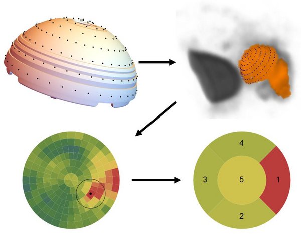
Fourteen patients receiving unilateral total hip arthroplasty were randomly assigned to receive a hydroxyapatite coated press fit cup (Trident) or a porous titanium coated hemispherical cup (Tritanium). For both groups migration (RSA) and biological activity (PET/CT) were measured at several follow-up moments. For the analyses of the PET/CT scans a new method was developed for visualisation of the 18F-fluoride uptake near the surface of the cup. The cup was separated into zones and measured activity was compared between cups, between zones and longitudinally.
Results
In several zones a significant difference (p<0.05) in activity was found between cup designs. For both designs higher activity could be found in the superolateral zones. Both designs showed a decrease in activity over time. RSA measurements did not show a difference in migration between the two groups. One patient was identified to have excessive migration of the cup and a pattern of bone formation that was different from the rest.
Conclusions
18F-fluoride PET/CT has the ability to perform measurements of the bone formation on the surface of the cup. These measurements can be used to compare cup designs and/or different regions of interest on the surface of the cup and can also be used for longitudinal studies.
Keywords
18F-fluoride PET/CT, Radiostereometric analysis, polar mapping, acetabular component