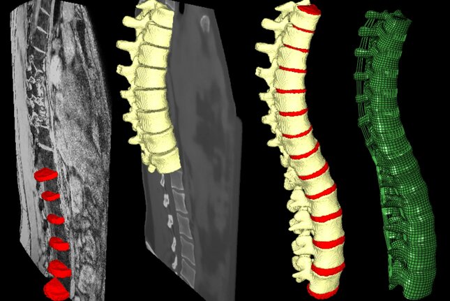
The generation of subject-specific finite element models of the spine is generally a time-consuming process based on computed tomography (CT) images. CT exposes patients and volunteers to harmful radiation and provides little information about soft tissues that play a crucial role in spine biomechanics. Through the use of artificial intelligence synthetic CT images can be generated from magnetic resonance (MR) images. This strategy provides information about both osseous and soft tissue structures while removing the required harmful radiation. Without radiation, it becomes possible to gather large amounts of 3D data of volunteers emphasizing the need for automation of morphological and biomechanical evaluations.
In this project, methods are developed for the automatic segmentation of MR and synthetic CT images, and for the generation of subject-specific finite element models. Deep-learning networks have been successfully trained for the automatic segmentation of the intervertebral disc, nucleus pulposus, facet joints, and vertebrae. For the automatic generation of finite element models aBayesian coherent point drift algorithm is employed to register a template mesh to the segmentations of the spine within a 1 mm accuracy. The segmentations can be used for 3D morphometric measurements and the mesh can be used for biomechanical evaluations, all completely automated.