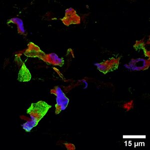
Bone is a highly dynamic tissue with multiple mechanical and metabolic functions that are maintained by the process of bone remodeling. In the bone remodeling cycle, progenitor cells are typically attracted and activated by biomechanical or biochemical signals after which resorption of mineralized bone matrix by monocyte derived osteoclasts starts. This phase is followed by a reversal phase in which osteoclasts leave, and towards osteoblasts differentiating mesenchymal stromal cells start populating the bone surface. These osteoblasts produce mainly collagen type 1 and embed themselves in their matrix over time. Once this matrix mineralizes, these osteoblasts differentiate towards osteocytes and obtain a more regulatory function in the remodeling process. In the healthy situation, bone resorption and formation are mostly in balance, resulting in no net bone loss or gain. In diseases such as osteoporosis and osteopetrosis, bone resorption and formation are typically unbalanced which can lead to respectively pathological bone loss or gain, affecting mainly bone’s mechanical functionality. Studies on these bone pathologies and their drug development are routinely performed in animal models. However, animal models represent human physiology insufficiently which is likely one of the reasons that only 9.6% of preclinically developed drugs are approved for regular clinical use. To enable the investigation of human healthy and pathological bone remodeling and to address the principal of reduction, refinement and replacement of animal experiments, the goal of this research is to develop a three-dimensional in vitro model for bone remodeling using a tissue engineering approach. In this model, osteoblasts, osteoclasts and osteocytes will be cultured together within one three-dimensional construct under physiological relevant biochemical and biomechanical stimuli. To study the interaction between these cells, (confocal) microscopy, scanning electron microscopy, micro-computed tomography, and biochemical analyses are performed.